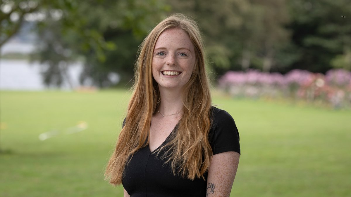
Emily Laura Meyer - PhD Scholarship 2025
Project summary:
Molecular Phenotyping of Arrhythmogenic Right Ventricular Cardiomyopathy by Spatial Proteomics
We study a specific heart disease where the main units of the heart, the heart muscle cells, become dysfunctional and are replaced by cells of fat and scar tissue. Using a precise laser method, we cut out individual cells from heart biopsies and analyze their contents. This allows us to see how each cell type changes in the diseased areas of the heart and helps us understand the processes driving the disease.
Project Title
Molecular Phenotyping of Arrhythmogenic Right Ventricular Cardiomyopathy by Spatial Proteomics
Background
ARVC is an inherited heart disease marked by loss of heart muscle and its replacement by fatty and scar tissue. This disrupts heart rhythm and can lead to sudden death, especially in young people. We still do not fully understand how different cell types interact in the diseased heart, limiting treatment development.
Aim
We aim to find out how heart muscle cells - as well as cells of fat and scar tissue - behave differently in hearts of patients with ARVC compared to healthy hearts. We aim to pinpoint disease-specific changes in each of these cell types and understand how their interactions contribute to scarring and fat buildup in the heart.
Methods
We use a method called laser microdissection to precisely cut out cells from heart tissue of ARVC patients and healthy individuals. These cells are then analyzed using a highly sensitive technique called mass spectrometry to examine their contents, specifically molecules called proteins. This lets us study how each cell type is affected by the disease while keeping their original location in the tissue in mind.
Preliminary results
Using this method, we identified around 1,600 proteins from as few as 10 laser-cut heart muscle cells, including proteins specifically found in the heart. This confirms the method’s sensitivity and specificity, showing we can robustly study individual cell types from heart biopsies.

Emily Laura Meyer
- MSc
- University of Copenhagen, Department of Biomedical Sciences
Main supervisor:
Alicia Lundby, Professor, Department of Biomedical Sciences, University of Copenhagen
Co-supervisor:
Henning Bungaard, Professor, Department of Cardiology, The Heart Centre, Rigshospitalet